Molecular and Microscopy Tools

Multicolor and 3D superresolution

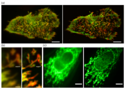
Multicolor imaging provides a powerful method for dissecting molecular relations and for tracking dynamic interplays (a-b). Fully 3D imaging remains a challenge in superresolution imaging. The robustness of pcSOFI and the inherent capacity of fluctuation imaging to deal with reduced signal-to-noise ratio allow us to achieve superresolution 3D imaging.

Multicolor and 3D Supperresolution

Activity biosensors reveal signaling dynamics in living systems

We developed several general strategies for engineering fluorescent biosensors to track the activities of second messengers, kinases and phosphatases. Quantitative measurement from live-cell fluorescence imaging can be combined with mechanistic computational modeling for systems analyses of signaling networks. We further demonstrated the first high-throughput compound screen based on reading dynamic kinase activities in living cells and are realizing live-animal imaging of dynamic signaling activities in collaboration with the Rick Huganir laboratory (JHU), extending the application of these fluorescent biosensors to chemical probe identification, drug discovery and mechanistic studies in en vivo contexts.

Research Graphic 1

Design of a Genetically Encoded Reporter for Imaging Kinase Activities

The genetically encoded Kinase Activity Reporter (KAR) contains a phosphoamino-acid-binding domain and kinase substrate sequence, sandwiched between a pair of fluorescent proteins that are capable of undergoing FRET. Phosphorylation of the substrate sequence by the kinase of interest results in the binding of the phosphoamino-acid-binding domain, induces a conformational change in the reporter that leads to an increase in FRET between the two fluorescent proteins.

Multicolor and 3D Supperresolution 2